Surface Anatomy Of Ribs / Https Fac Ksu Edu Sa Sites Default Files Anatomy Lecture 19 Ul Pdf : Atypical ribs rib 1 is shorter, most curved and wider than the other ribs.
Surface Anatomy Of Ribs / Https Fac Ksu Edu Sa Sites Default Files Anatomy Lecture 19 Ul Pdf : Atypical ribs rib 1 is shorter, most curved and wider than the other ribs.. Surface anatomy of the human body, front. The ribs are elastic arches of bone, which form a large part of the thoracic skeleton. We think this is the most useful anatomy picture that you need. In most tetrapods, ribs surround the chest, enabling the lungs to expand and thus facilitate breathing by expanding the chest cavity. The second rib articulates with the sternum at the sternal angle, making this site an excellent landmark for determining rib number.
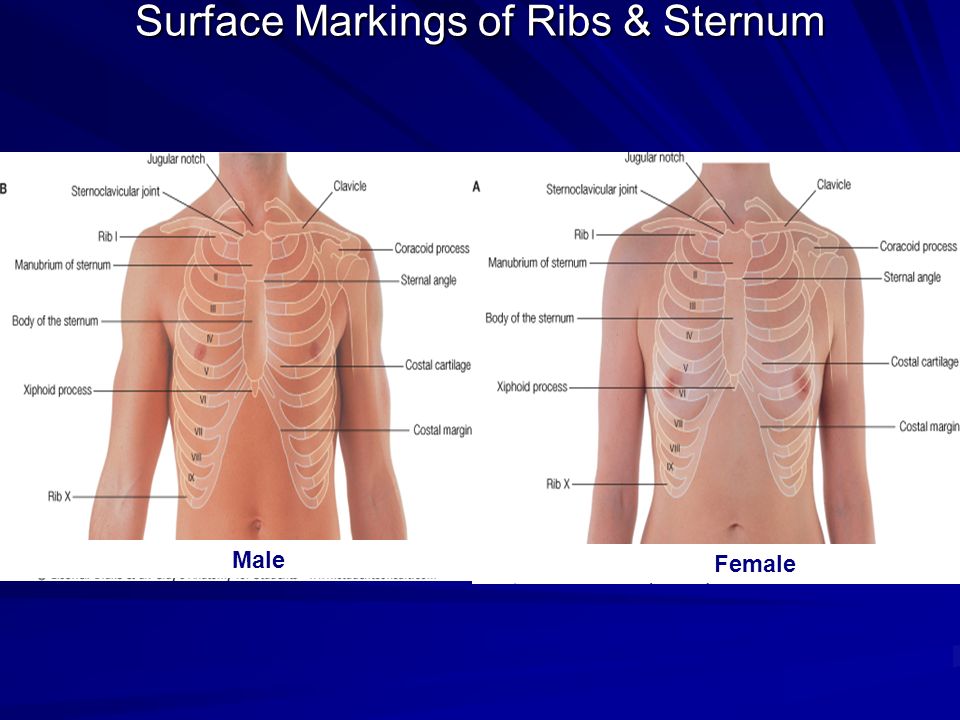
Colour atlas of human anatomy volume 1, 6th edition, trunk, ribs, pg. Ascending aorta ends and arch of aorta begins. Bony landmarks.—the second costal cartilage corresponding to the sternal angle is so readily found that it is used as a the influence of the obliquity of the ribs on horizontal levels in the thorax is well shown by the following line. The rib cage forms the majority of the thoracic skeleton. Learn vocabulary, terms and more with flashcards, games and other study tools.
1 Pratt Lung Surface Anatomy Flashcards Quizlet from quizlet.com Superficial dissection of the back of the neck. Ascending aorta ends and arch of aorta begins. Learn the true ribs, false ribs, and floating ribs, as well as the difference between in this anatomy lesson, i'm going to cover the rib bones, also called costae in latin. In vertebrate anatomy, ribs (latin: Bony landmarks.—the second costal cartilage corresponding to the sternal angle is so readily found that it is used as a the influence of the obliquity of the ribs on horizontal levels in the thorax is well shown by the following line. The ribs are elastic arches of bone, which form a large part of the thoracic skeleton. There are two types of ribs, namely typical and atypical. Surface markings of the thorax.
In vertebrate anatomy, ribs (latin:
They extend from the inner surface of one rib to the inner surface of either the next rib or even the one below that. Surface anatomy of the back. The ribs form the main structure of the thoracic cage protecting the thoracic organs, however their main function is to aid respiration3. They are twelve in number on either side; An exception to this rule is those closest to the skin's surface run from the back of the vertebrae to the scapula eg trapezius , rhomboid s, latissimus dorsi , others wrap around the. The surface anatomy of the ear is frequently cut and reconstructed during mohs surgery. Rib fractures usually result from blows or from crushing injuries. Surface anatomy (also called superficial anatomy and visual anatomy) is the study of the external features of the body of an animal.1 in birds this is termed topography. Now notice the rib belongs to the side on which it is both ends touch the surface. It can help you understand our world more detailed and specific. The exceptions are the 11th and 12th ribs that don't have this surface, which enables them much higher mobility. Surface anatomy of the human body, front. The ribs stretches posteriorly from thoracic vertebrae to the anterior lateral edges of the sternum.
Lie on top of pectoralis major and tail extends articulation with costal cartilage of rib ii. Now notice the rib belongs to the side on which it is both ends touch the surface. Superficial dissection of the back of the neck. There are twelve pairs of ribs, all of which articulate with the vertebral column, while only the first seven ribs directly articulate with the sternum. They extend from the inner surface of one rib to the inner surface of either the next rib or even the one below that.
Thorax Thoracic Wall Muscles Of Respiration Ppt Video Online Download from slideplayer.com Surface anatomy „ four techniques when examining surface anatomy „ visual inspection „ directly observe the structure and markings of surface sternal angle is clinically important because it is at the level of the costal cartilage of the second rib. They are twelve in number on either side; The loose segment of the wall moves. Rib fractures usually result from blows or from crushing injuries. Each rib articulates posteriorly with two thoracic vertebrae by the costovertebral joint. This muscle assists the internal intercostal muscles. Surface anatomy of the back. Anatomy ▶ thorax ▶ bones and cartilages ▶ the ribs.
An exception to this rule is those closest to the skin's surface run from the back of the vertebrae to the scapula eg trapezius , rhomboid s, latissimus dorsi , others wrap around the.
Surface anatomy (also called superficial anatomy and visual anatomy) is the study of the external features of the body of an animal.1 in birds this is termed topography. Lie on top of pectoralis major and tail extends articulation with costal cartilage of rib ii. Surface markings of the thorax. (subclavian means below the clavicle. Gross anatomy there are 12 pairs of ribs which are separated by intercostal spaces. This image added by anatomy is the amazing science. The ribs stretches posteriorly from thoracic vertebrae to the anterior lateral edges of the sternum. You can click the image to magnify if you cannot see clearly. Superior mediastinum separated from inferior. Learn vocabulary, terms and more with flashcards, games and other study tools. The rib cage is made up of 12 pairs of ribs, each having a posterolateral bony and an anterior costal cartilaginous component ( fig 4.2 ). This muscle assists the internal intercostal muscles. Muscles.—the surface muscles covering the thorax belong to the musculature of the upper extremity (figs.
The ribs help protect vital organs in the thorax such as the heart. The channel provides a pathway. This muscle assists the internal intercostal muscles. Surface markings of the thorax. Surface anatomy and surface markings bibliographic record list of illustrations subject index.
A B Surface Projections Of The Pleurae And Lungs Download Scientific Diagram from www.researchgate.net We think this is the most useful anatomy picture that you need. Some have everyday names like the palm of the hand , the sole of the foot , and the nape of the neck. There are twelve pairs of ribs. Landmarks of the thoracic wall. This image added by anatomy is the amazing science. Costae) are long, flat, curved bones that form the rib cage. We hope you will use this picture in the study and. Anatomy of the human body.
There are twelve pairs of ribs.
The surface regions of the body have received their names in a variety of ways. They extend from the inner surface of one rib to the inner surface of either the next rib or even the one below that. The ribs help protect vital organs in the thorax such as the heart. Right and left scapular li. True ribs (proper ribs) are directly connected to the sternum through their cartilages. 91215, 91219), and will be described in that section (page 1325). The rib cage is made up of 12 pairs of ribs, each having a posterolateral bony and an anterior costal cartilaginous component ( fig 4.2 ). Ascending aorta ends and arch of aorta begins. In vertebrate anatomy, ribs (latin: Superficial dissection of the back of the neck. The surface anatomy of the ear is frequently cut and reconstructed during mohs surgery. Anterior chest between ribs i and vi & between the sternum and anterior axillary line. If the rib is set on the incorrect side, then only its anterior end.
Costae) are the long curved bones which form the rib cage, part of the axial skeleton anatomy of ribs. Surface markings of the thorax.


Posting Komentar
0 Komentar